
Microscopic Enhanced Dentistry
Surgical Microscopes
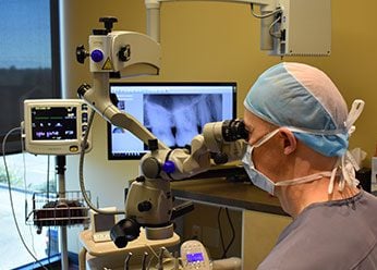
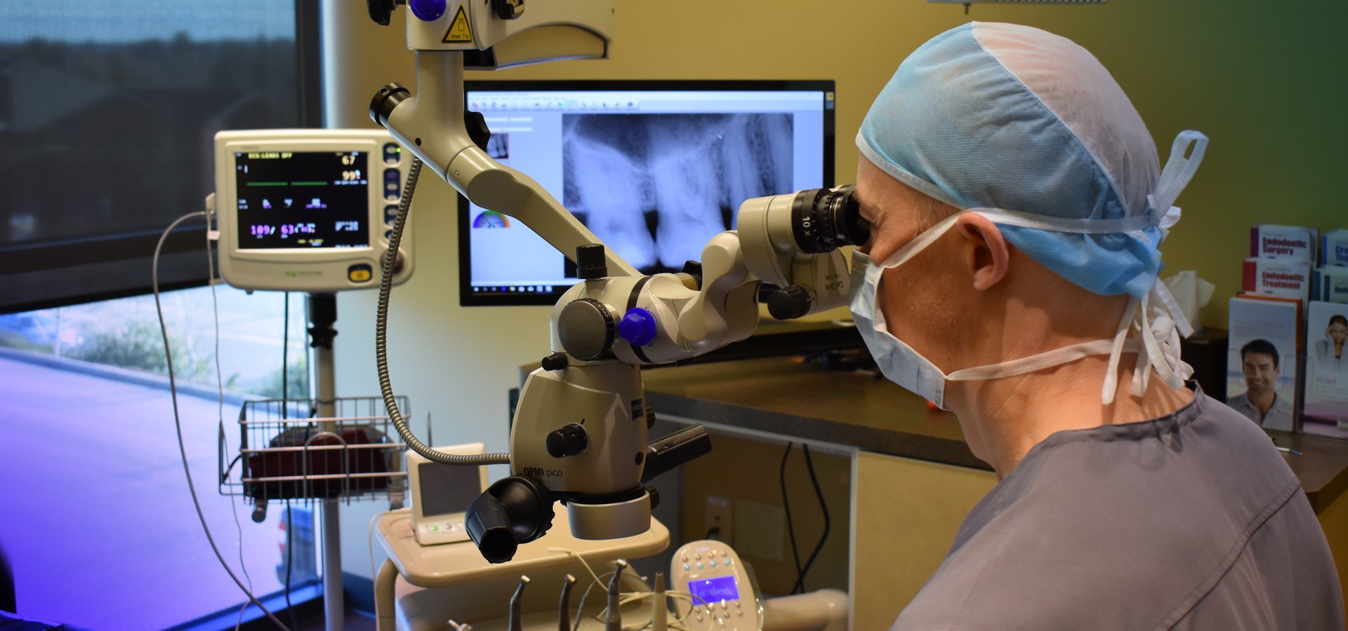
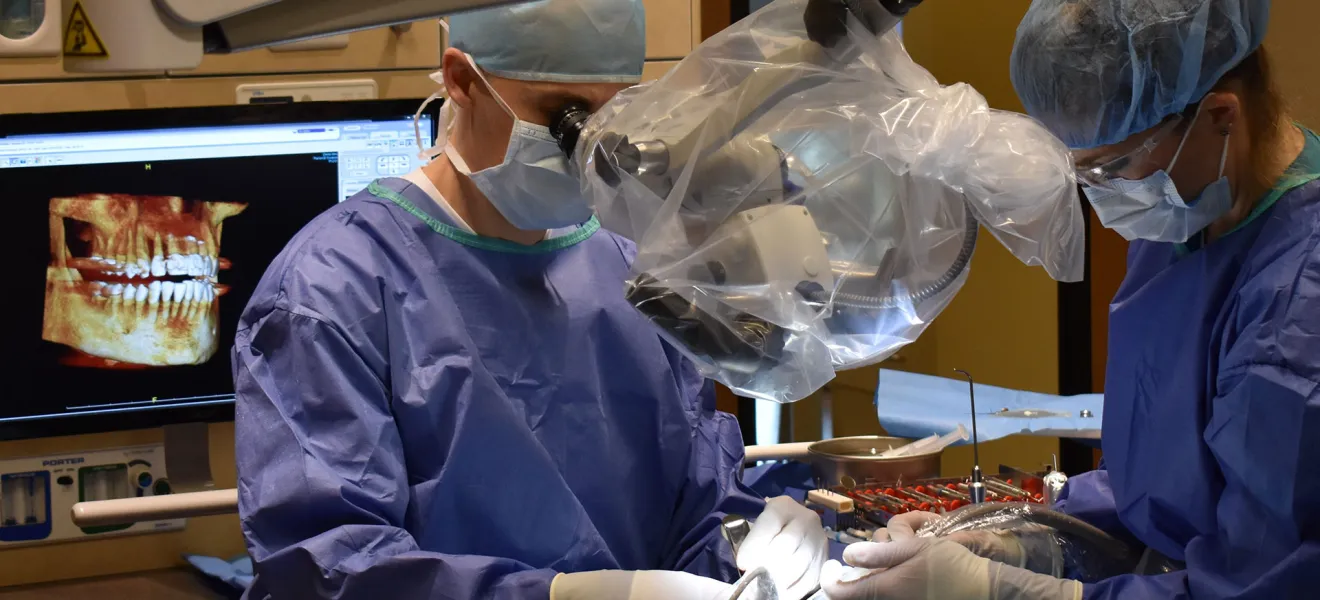
The introduction of the surgical microscope in dentistry has revolutionized the field of root canal therapy, endodontic microsurgery, bone & soft tissue grafting and implant therapy. We have invested in the very best quality surgical microscopes, by Carl Zeiss, that provide unparalleled magnification and illumination for all of our surgical and non-surgical procedures.
The operating microscope, which was first introduced to dentistry in 1981, offers significantly increased magnification and illumination of the operating field compared to the naked eye and even magnifying loupes “glasses” with attached headlamps. The improved vision allows for dramatically increased precision in the clinical practice of dentistry. Even what we consider the simplest of dental procedures involves being able to see the minutest of detail. In regards to dentistry, “if we can see it, then we have the opportunity to treat it”. Microscopic examination and microsurgical techniques have transformed the way the modern clinician practices.
The operating microscope has had a significant impact on the quality of dental care since it’s inception. The specialty of Endodontics has seen the most rapid incorporation of the operating microscope into dental practice. With root canal therapy numerous studies over the past decade have shown the microscope’s advantages in locating the internal anatomy of teeth, detecting cracks, and in performing root-end microsurgery. With surgical treatment the enhanced vision through the microscope allows for more precise treatment. The microscope allows for increased precision with surgical incisions, suturing of soft tissue, placement of bone and tissue grafts, treating defects of the teeth, and surgical placement of implants. With the aid of the microscope many advanced surgical procedures such as bone and tissue grafts can be performed through the site of an extracted tooth without having to elevate a tissue flap. Even with restorative dental treatment, the enhanced vision allows for increased precision for removal of decay and restoring teeth with crowns and fillings.
My first introduction to the operating microscope was during my training in dental school. The first time I looked through the microscope I realized what I previously was unable to see without it. I realized the significance this one technology had on being able to provide improved dental therapy. Today I utilize the microscope in all phases of my practice from performing micro-root canal therapy to utilizing microsurgical techniques with bone, soft tissue and implant therapy. Dr. B. Wilson


